Chemical or Physical Agents which are used to prevent and preserve the tissue are called fixatives.
Types of Fixatives:
Physical Agents:
- Heat
- Ultra Violet Rays
- Microwaving
- freeze-drying
Chemical Agents:
- Coagulants
- Non Coagulants
Classification of Fixative:
On the basis of working condition
- 95% ethyl alcohol best fixative for cytology
- Formalin best fixative for histopathology
Hopwood Classification of Fixatives:
1. Aldehyde Fixatives:
- Formaldehyde Fixatives
- Gluteraldehyde Fixatives
- Acrolein
- Glyoxal
2. Oxidizing Agents:
- Osmium tetroxide
- Potassium permanganate
- Potassium dichromate
3. Protein Denaturing Agents:
- Acetic Acid
- Methyl Alcohol
- Ethyl Alcohol
4. Cross Linking Agents:
- Carbodiimides
- Dimethyl Subrimidate
5. Unknown Mechanism/Miscellaneous:
- Mercuric Chloride
- Picric Acid (Explosive when dry or when complexes with metal and metallic salts)
Aldehydes:
- Aldehydes include formaldehyde (formalin) and gluteraldehyde.
- Tissue is fixed by cross-linkages formed between the proteins particularly between lysine residues.
- Only those lysine residues which are on the exterior of protein molecule react.
- The reaction with formaldehyde is largely reversible by excess of water especially over the first 24 hour of reaction time.
- With gluteraldehyde the reaction is rapid and irreversible.
- Reaction between aldehydes and proteins is pH dependent proceeding more rapidly at high pH.
- Fixation denatures proteins to some extent which is acceptable and satisfactory for routine pathology work.
- For enzyme histochemistry, immunocytochemistry and electron microscopy this issue should be considered.
- Enzyme molecules and antigenic sites must not be altered or destroyed by chemical fixation.
- Shapes of DNA and RNA should not be changed.
- Gluteraldehyde causes loss of up to 30% of αhelix structure of protein and formaldehyde markedly less.
- Formaldehyde does not give as good morphological picture as gluteraldehyde.
- Cross linkage does not damage the arrangement of proteins significantly therefore antigenicity is not vanished.
- Therefore, formaldehyde is good for immunoperoxidase techniques. Formalin penetrates tissue well, but is relatively slow.
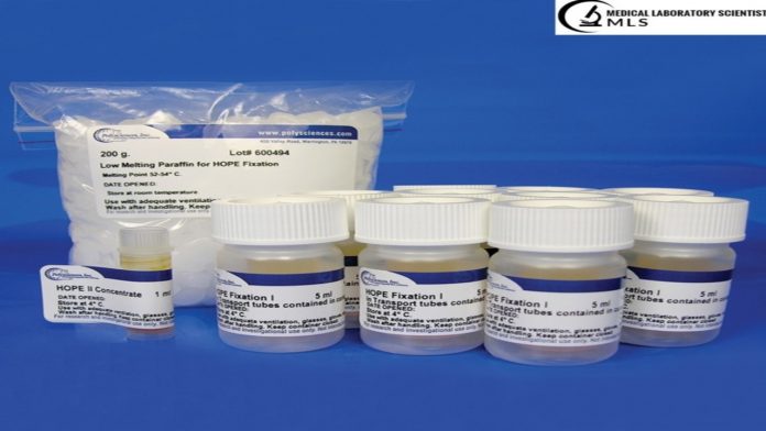
- Normal solution is about ten (10%) percent neutral with buffered formalin solution.
- A buffer stops acidity which stimulate autolysis and reason for precipitation of formol haem pigment in the tissues.
- Formaldehyde in its 10% neutral buffered form.
- Neutral Buffered Formalin (NBF) is the best fixative in diagnostic pathology labs.
- Pure formaldehyde is a vapor which when completely dissolved in water forms a solution containing 37–40% formaldehyde; this aqueous solution is known as ‘formalin’.
- The typical 10% formalin used in the fixation of different histopathological. This may consists of four (4%) percent weight/volume of formal.
Formaldehyde Fixatives:
- 37% Formaldehyde is available commercially and should be stored at room temperature in close and air tight container
- Clear, colorless and no precipitates
- Storage should not be more than 1 year
- Already opened bottles should not be used more than six months
- 10% formalin should not be used more than 3 months.
Other variants of formaldehyde containing fixatives:
- Mostly in lab formaldehyde buffered to pH 7 with phosphate buffer is used.
- Use of neutral buffered formalin stops formation of formalin pigment which forms in non-buffered formaldehyde solutions.
- For lipids formol calcium is used.
- Alcoholic formalin has the advantage of rapid fixation and especially recommended for glycogen.
- They are carcinogenic.
- PPE should be wear during working with formalin.
Gluteraldehyde:
- Gluteraldehyde causes deformation of alpha-helix structure in proteins so it is not good for immunoperoxidase staining.
- Though, it fixes very rapidly and excellent for electron microscopy.
- It infiltrates/penetrate very poorly and unwell, but provides finest cytoplasmic and nuclear features.
- The standard solution is a 2% buffered Gluteraldehyde
- Rapid irreversible reaction have led to its use as a sterilizing agents of fiberoptic endoscope
Mercuric Chloride Containing Fixative:
- These fix tissue by an unknown mechanism.
- Examples include fixatives as Zenker, Susa and Helly’s fluid.
- These fixatives penetrate poorly and cause some tissue shrinkage so mostly used in combination of other fixatives or as secondary fixatives.
- Their best application is for fixation of hematopoietic and reticuloendothelial tissues and fetal brain
- They comprise mercury so should be dispose of sensibly.
- Mercuric chloride is significantly preferred for its abilities of improving the staining qualities of tissues, mostly for trichrome stains.
- Tissues fixed with mercuric chloride fixatives (except susa) contain black precipitate
- This is removed by treating in alcoholic iodine soln. and then decolorizing in sodium thiosulfate.
Alcohol Containing Fixative:
- Alcohols, as well as methanol and ethanol, are protein denaturants and are not suitable for daily basis for tissues because they create too much brittleness/fragility and hardness/rigidity in the tissues.
- They are excellent for cytological smears because their action is rapid and provide maximum nuclear details and features.
- Spray cans of alcohol fixatives are promoted to labs perform PAP smears but inexpensive hairsprays do fair as well.
- Recommended for preservation of glycogen
- Example include carnoy’s fixative
Oxidizing Agents Fixatives:
- Oxidizing mediators/agents contain permanganate fixatives for example potassium permanganate, dichromate fixatives such as potassium dichromate, and osmium tetroxide.
- Few of them have specific applications but are used very rarely.
- Some of them are identified according to their appliance of action but osmium tetroxide make crosslinking with proteins.
Picric Acid Containing Fixative:
- They have an unknown mechanism of action.
- Examples include Bouin’s, gendre’s and rossman’s fluid.
- Picric acid has ability of burst/explosive danger in dry its form.
- Yellow color may be removed but another acid dye or lithium carbonate or washing with changes of 50-70% alcohol
Reaction of fixatives of proteins:
- In fixation of tissues for routine histopathology the most important reactions are those which stabilize proteins
- Proteins comprise more than 50% of dry body weight
- The principle involved is that the fixative have the property of forming crosslinks with proteins forming a gel which provides strength and keeping everything as close to their in vivo state as possible
- Soluble(globular) proteins are fixed to structural(fibrous) proteins and rendered insoluble and whole structure is given mechanical strength allowing subsequent preparations to take place
- Fixation normally takes place in aqueous solution. Occasionally vapors can be used.
Reaction of fixatives with Nucleic Acids:
- Fixation bring about changes in physical and chemical state of DNA & RNA
- In their native state DNA & RNA do not react with formaldehyde at room temperature
- Reaction mixture are heated 450C for RNA and 650C for DNA
- High temperature relates to the uncoiling of DNA & RNA
- Up to 30% of nucleic acids are lost during fixation
- Alcoholic fixatives like ethanol, methanol and carnoy’s fixative are commonly used
- DNA will collapse at 65% conc. Of ethanol
Reaction of fixatives with Lipids:
- They are largely removed during tissue preparation
- If lipid are to be demonstrated specifically frozen sections or cryostat should be used
- Formol calcium is the choice of fixative
Reaction of fixatives with Carbohydrates:
- Alcoholic fixatives are the fixative of choice for glycogen.




















Thank you so much, I really appreciate…
Thank you so much
gr8