Urinalysis: Chemical Examination of Protein, Sugar, Bile Pigments & Bile Salts
In Urinalysis, Urine chemical examination is a fundamental component of a urinalysis, a diagnostic test that assesses the physical and chemical properties of urine. This analysis provides valuable information about a person’s overall health, helps diagnose various medical conditions, and monitors the progress of certain diseases.
PROTEINS IN URINE:
Normal Levels: In normal urine, proteins are typically undetectable by routine methods. Their presence can be a significant indicator of renal diseases and may be used to monitor therapy in such conditions.
Conditions Associated with Proteinuria: Proteinuria (presence of proteins in urine) can occur in various conditions, including hypertension, pre-eclamptic toxaemia, renal parenchymal diseases, and urinary tract infections.
METHODS FOR MEASURING PROTEINS:
1. Turbidimetric Method
Heat Method
This involves heating a urine sample, which coagulates proteins (mainly albumin), similar to how boiling coagulates egg white. Adding acetic acid can precipitate albumin, globulins, and proteoses, causing turbidity.
Acid Precipitation
Chemical agents like sulfosalicylic acid and nitric acid can be used to precipitate proteins. However, these agents may also precipitate other urinary constituents.
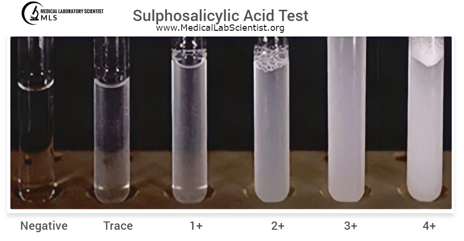
Sulfosalicylic Acid Test (Kingsbury and Clark)
This test relies on denaturing and precipitating proteins with acids. A urine sample is mixed with sulfosalicylic acid, and turbidity is compared to standards. False positives can occur due to mucous, iodine contrast media, metabolites of tolbutamide, plasma expanders, IV albumin, and sulfisoxazole. X-ray contrast media can also lead to false positives for up to three days after administration. Highly buffered urine may produce false negatives.
2. Colorimetric Method
At pH 3.0, tetrabromophenol blue is yellow in the absence of protein but turns green to blue in the presence of protein. This color change is dependent on the protein concentration. Various reagents, including sulfosalicylic acid, citrate buffer, nitric acid, and tetrabromophenol blue, can be applied to test strips for this purpose. Trichloracetic acid with Exton’s reagent is another option. These tests are highly sensitive and can detect proteins in the range of 0.05-0.2g/L. However, confirmatory testing with the turbidimetric method is recommended. The test is specific for albumin, and false positives can occur in alkaline or highly buffered urine. Improper color matching or lighting conditions can also lead to false results.
GLUCOSE AND REDUCING SUGARS:
Principle
Monosaccharide hexoses are considered reducing sugars. They produce color reactions when tested with Benedict’s reagent or clinitest tablets.
- Hexose Sugars: Monosaccharide hexoses such as glucose, galactose, and fructose are reducing sugars.
- Polysaccharides: Naturally occurring polysaccharides like glycogen (found in animal tissue) and starch (found in plants) are long-chain carbohydrates composed of glucose subunits. Glycogen is highly branched, while starch consists of both straight chains (amylose) and branched chains (amylopectin).
- Testing: Benedict’s reagent and clinitest tablets are commonly used to detect reducing sugars in urine, with the appearance of a color indicating their presence.
- Specificity: These tests are sensitive to a range of reducing sugars, including glucose. However, they are not specific to glucose and can detect other reducing sugars as well.
- Clinical Significance: Detecting glucose in urine can be indicative of conditions like diabetes mellitus, where elevated blood glucose levels can spill into the urine. It is a valuable diagnostic tool for monitoring and managing diabetes.
Common Reducing and Non-Reducing Sugars:
|
|
Reducing Sugars |
Non-Reducing Sugars |
|
Monosaccharides |
Glucose Fructose Galactose |
|
|
Diasaccharides |
Lactose (galactose+glucose |
Sucrose (fructose+glucose) |
|
|
Maltose (glucose+glucose) |
|
GLUCOSE AND REDUCING SUBSTANCES IN URINE:
- Normal Glucose Excretion: In a healthy adult, the normal excretion of glucose in urine is minimal, typically up to 130 mg/24 hours.
- Reducing Sugars: While glucose is the most common reducing sugar found in urine, other reducing substances can also be present (see Table 9.4).
- Excess Glucose in Urine: Elevated levels of glucose in urine can occur in conditions such as diabetes mellitus, renal glycosuria, post-gastrectomy, epinephrine excess (from adrenal glands or therapeutic injections), pancreatitis, hyperthyroidism, liver damage, renal tubular disease, after a heavy meal, and due to emotional stress.
Benedict’s Test:
Benedict’s test is a non-specific method used to screen urine for the presence of reducing substances. A similar test is incorporated into Clinitest tablets.
Principle
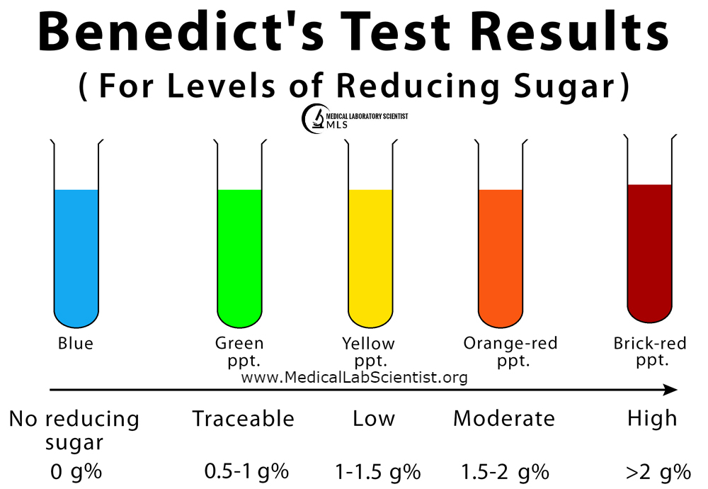
Benedict’s test relies on the reduction of soluble blue cupric ions (CuSO4) in a heated, strongly alkaline solution. Urinary reducing agents reduce these cupric ions to form yellow-red insoluble cuprous ions (Cu2O). The change in color from blue to orange-red indicates a positive test.
Procedure
- Pour 5 ml of Benedict’s reagent into a test tube.
- Heat the reagent to exclude false positive results.
- Add 0.5 ml of urine to the test tube.
- Boil the mixture for an additional 2 minutes.
- Cool the test tube under running tap water.
- Observe the color of the precipitate.
Interpretation
The results can be interpreted based on the color change, as indicated in Table 9.3. This method is sensitive and can detect glucose at concentrations as low as 0.2%. It is also positive for other reducing substances listed in Table 9.4. Additionally, it may yield positive results with substances like salicylates, chloral hydrate, formalin, Vitamin C, and certain drug metabolites such as nalidixic acid and first-generation cephalosporins.
Key Points
- Benedict’s test screens for the presence of reducing substances in urine.
- It is a non-specific method sensitive to glucose and other reducing agents.
- Positive results are indicated by a color change from blue to orange-red.
- Interpretation is based on the color of the precipitate.
- The test can detect glucose at low concentrations and is used to screen for conditions such as diabetes mellitus and renal glycosuria.
- Positive results may also occur with other substances, so further confirmatory testing may be needed for a precise diagnosis.
Reducing substances in urine that may give a positive reaction with Clinitest tablet/Benedict’s test:
|
Reducing substance |
Comment |
|
Glucose Glucoronates |
Common |
|
Lactose |
Common in pregnancy |
|
Galactose Fructose Pentose Homogentisic acid |
Rare |
|
Urate Creatinine |
Weakly positive at high concentrations |
ENZYMATIC TEST FOR GLUCOSE:
Specific for Glucose
This test is specifically designed to detect glucose and is now available on dipsticks. It relies on the enzymatic reaction in which glucose is converted to gluconic acid and hydrogen peroxide (H2O2) by glucose oxidase in the presence of oxygen. The produced H2O2 reacts with orthotoluidine in the presence of peroxidase, resulting in the formation of colored compounds.
Sensitivity
The enzymatic glucose test is highly sensitive and can detect glucose at concentrations as low as 0.1%. It provides a specific assessment of glucose levels in urine.
False Results
This test is not affected by normal urine constituents and does not yield false-negative or false-positive results. However, it may produce false positives in the presence of bleach and peroxides used for cleaning containers. Very high doses of Vitamin C and homogentisic acid may lead to false negatives.
Precautions
When using test strips, it’s important to follow the precautions provided by the manufacturers.
Interpretation
A positive result with Benedict’s test and a negative result with the enzymatic glucose test may suggest the presence of non-glucose reducing substances, such as galactose, pentose, or lactose.
- Galactose
Indicates Galactosaemia: The presence of galactose in urine indicates galactosaemia, an inborn error of carbohydrate metabolism. This condition arises due to a deficiency of galactose-1-phosphate uridyltransferase, which normally converts galactose to glucose-1-phosphate in the liver. Galactosaemia is not common and typically presents in infancy. Infants with this condition may develop cataracts, liver damage, and potential mental retardation.
- Pentose
Indicates Pentosuria: Pentosuria is an inborn error of metabolism characterized by the excretion of pentose-L-xylulose in urine. Pentosuria can also occur after the ingestion of raw plums or cherries. It can be checked using the bial-orcinol test.
- Lactose
Occurrence: Lactose, another sugar, may be found in urine during late pregnancy, lactation, or in individuals with extremely high milk diets.
- Lactose Intolerance
Lactose intolerance may result in lactosuria, which is a rare metabolic condition associated with the inability to properly metabolize lactose, leading to its presence in urine.
BILE PIGMENTS (BILIRUBIN):
Purpose: Testing for bilirubin in urine is essential for screening, diagnosing, and monitoring liver, biliary, and hemolytic diseases.
Normal Levels: Normally, urine bilirubin levels are less than 0.03 mg/dl and are undetectable by routine tests.
Conditions: Bilirubin may be found in urine in conditions such as cirrhosis of the liver, viral hepatitis, carcinoma of the head of the pancreas, bile duct obstructions, and hemolysis.
METHODS FOR DETECTING BILIRUBIN:
Foam Test: This outdated method involved shaking a urine specimen and observing the color of the foam. It was insensitive and is no longer used.
Dye Dilution Test
This method, using methylene blue, was also insensitive and is obsolete.
Fouchet’s Test
Barium chloride is used to precipitate phosphates and concentrate bile pigments, which are then tested for using the oxidation reaction. The sensitivity of this test ranges from 0.005 to 1.0 mg/dl. False positives can occur with salicylates, but the color produced is purple. Pyridium-like substances (e.g., phenazopyridine) can give a red color.
Diazotization Test
In this test, a stabilized diazo compound reacts with bilirubin to form a blue color. This test is specific and can detect bilirubin at concentrations as low as 0.05 mg/dl. It is sensitive and should be performed on fresh urine. Very large amounts of phenothiazine (chlorpromazine) metabolites can yield false positive results, and the presence of pyridium-like substances can give a red color.
BILE SALTS:
Hay’s Test: This test relies on the principle that bile salts lower surface tension, causing light powdered sulfur to sink to the bottom.
Detection of Bile Salts
- Procedure: To detect the presence of bile salts in urine, take 5 ml of urine in a test tube. Sprinkle a small amount of finely powdered sulfur granules on the surface of the urine. If the sulfur granules sink, it indicates the presence of bile salts in the urine.
- Caution: False-positive results may occur due to the sinking of heavy impurities in the sulfur powder.


























[…] Urinalysis: Chemical Examination of Protein, Sugar, Bile Pigments & Bile Salts […]